Amazing Microscope Images taken by Two UC Chile Students Surprise Scientists
Two doctoral students from the Biological Sciences Faculty were recognised for their unique microscopic images showing a group of rat sensory neurons. Whilst one of the images was selected by Nature Magazine, the Chilean Society for Cell Biology chose the other
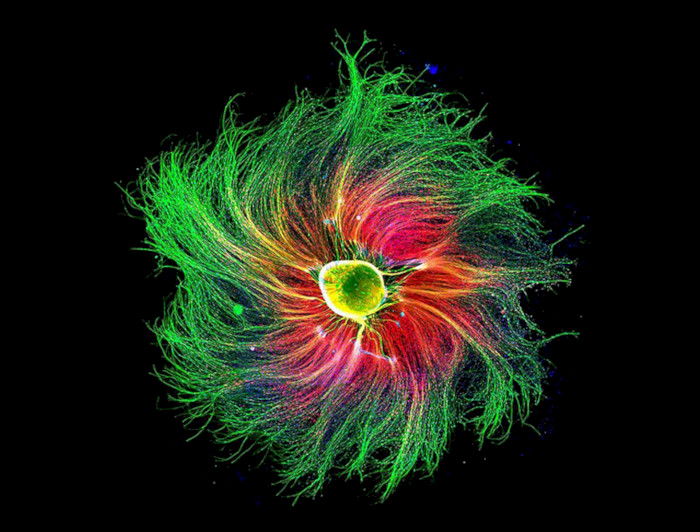
photo_camera The colorful image of a rat embryo’s dorsal root ganglion, a group of sensory nerve cells that detect pain.Paula Díaz Céspedes.
Two doctoral students from the Faculty of Biological Sciences were recognized for their unique microscopic images showing a group of rat sensory neurons. Whilst one of the images was selected by Nature Magazine, the Chilean Society chose the other for Cell Biology.
A photo of a group of rat sensory neurons was the only Chilean image selected by Nature Magazine for its theme "The best science images of 2021".
Paula Díaz Céspedes took the microscopy photo. She's a Thesis student in a Doctoral Program in Physiological Sciences at the Faculty of Biological Sciences and the Millennium Nucleus for the Study of Pain (MiNuSPain).
To capture the image, Paula Diaz used a fluorescence microscope—which allows observation with fluorescent contrast— to obtain the colorful image of a rat embryo’s dorsal root ganglion, a group of sensory nerve cells that detect pain, among other things.
"I think I was selected because pain is a prevalent condition with psychological and social impact. This model allows us to study pain, and we can see the green fibers that are the axons, which in mammals can grow to meters in length," said the student.
It makes sense to Paula that the image—which had previously won Fourth Place in the Nikon's Small World Photomicrography Competition (2021)— was selected when Nature magazine was looking to include alternative topics besides COVID in 2021.
The topics ranged from climate change and studies of genetic diseases, to black holes and even neuroscience.
"I don't think these images are foreign to Covid. There is a correlation between climate change and the pandemic as these are events shaped by biological, psychological and social factors, same as pain."
Currently, Paula Díaz is undertaking a doctoral internship abroad for the National Research and Development Agency (ANID) in the University of Oxford's Nuffield Department of Clinical Neurosciences. Her studies aim to learn how to identify human stem cells' sensory neurons to develop pain transferal research in Chile.
The student is a thesis student of MiNuSPain, which aims to advance knowledge of the pathophysiology of pain, contribute to the formation of advanced human capital in biomedical and clinical sciences, and share scientific knowledge.
"Pain and its many classifications is a disease. I am interested in studying neuropathic pain because it is key to unify criteria to establish standardized parameters to validate pain research. This is why getting the right diagnosis is key as it will lead to the adequate treatment,"
Close-up of a Fly Larva's Brain
Francisca Rojo, a fellow student of the Faculty of Biological Sciences, also won an award by the Chilean Society for Cell Biology for the best image in Cellular and Molecular Biology 2021.
The award takes place during the Annual Meeting of the Chilean Society for Cell Biology, where many scientists from other faculties and universities participate.
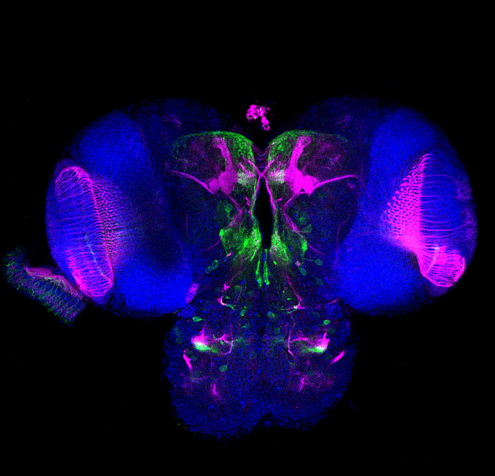
The image titled "Expression of Lipophorin Receptor 1 (LpR1) in D. melanogaster fly larva brain" was obtained using the Zeiss Airsycan microscope of the Advanced Microscopy Unit.
The larval brain of a Drosophila melanogaster, or fruit fly, measures approximately 0.5 mm. Francisca's challenge was to remove the brain and dissect it without damaging it, a task that took her a couple of years to achieve.
This photo was taken during the pandemic, between quarantines, and was part of the final experiments for a scientific publication currently under review.
According to Francisca Rojo:
"The photo is a representation of the larval brain of the fly Drosophila melanogaster:
- The brain areas where LpR1 is expressed are marked in green.
- Fasciclin II, a protein highly expressed in the fungiform body of the fly, is seen in magenta, which allows visualisation. The fungiform body is a brain structure active in several insects, whose primary known function is the processing and storing of olfactory memory.
- The blue mark is staining with Dapi, a fluorescent chemical compound that binds to the DNA, allowing cell nuclei to be marked."
Francisca is a Biological Sciences PhD student in Cellular and Molecular Biology. She is working on her thesis under the supervision of professors María Paz Marzolo and Jorge Campusano, in two laboratories of the faculty:
- the Intracellular Traffic and Signaling lab
- the Behavioral Neurogenetics lab
Today she is in France doing a research internship in the laboratory of Dr. Serge Birman at the École Supérieure de Physique et de Chimie Industrielles de la Ville de Paris (ESPCI-PLA).
During the internship, she hopes to obtain results that will allow her to advance her doctoral thesis and contribute to her host laboratory.
"My thesis seeks to describe the functions that Lipophorin receptors have on the structure and function of the D. melanogaster fungal body, beyond the role of lipid transportation, which is why this family of well-known proteins."
"Furthermore, from studies in mammals, there is evidence that the homologous proteins present in these organisms take part in the correct development of brain structures, helping to position the neurons correctly. In more adult stages, they could have a role in neuronal plasticity. In my thesis, I am trying to determine if these functions are preserved in the fly," explained Francisca.


