Fly Brain Research Provides Insight into Diseases of the Human Brain
A groundbreaking study led by PhD in Biological Sciences, Francisca Rojo, could be key to understanding the development of brain structures and provide new insights into diseases such as schizophrenia and neurodegenerative diseases associated with ageing.
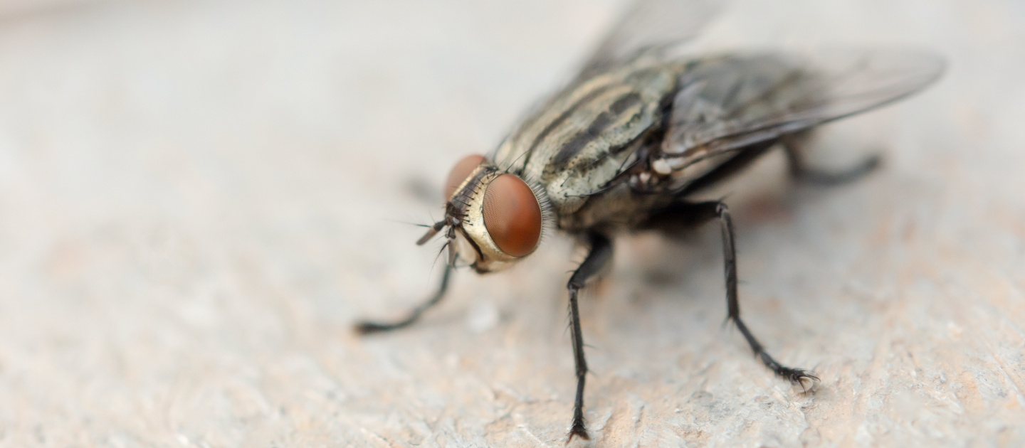
photo_camera Francisca Rojo's interdisciplinary research, which was published in the prestigious journal BMC Biology, advances our understanding of how molecular signals drive the formation of a brain structure. (Photo by: iStock Photo)
We have been studying flies to answer crucial biology questions for more than 100 years.
Did you know that the principles of how genes contribute to embryonic development were discovered through studies of fruit flies? In fact, that discovery earned Professors Edward Lewis, Christiane Nüsslein-Volhard, and Eric Wieschaus the Nobel Prize in Physiology and Medicine in 1995.
Flies are popular research subjects amongst biologists around the world, including in Chile, specifically at the Faculty of Biological Sciences of UC Chile.
Recently, a team from the “Intracellular Traffic and Signaling” and “Behavioral Neurogenetics” laboratories, both from the Department of Cellular and Molecular Biology, discovered something extraordinary while investigating the fruit fly (Drosophila). They found that certain types of brain proteins known as lipophorins are not only able to transport lipids, but are also key to the development of brain structures and the correct positioning of neurons.
Their paper, which was published in the scientific journal BMC Biology, describes the functions of these proteins beyond a role in lipid transport (which is what lipophorins are known for in the scientific literature).
The discovery has attracted international interest because, until now, these proteins have only been studied in insects in the context of lipid transport disorders.
As Francisca Rojo, PhD in Biological Sciences, specialized in Cellular and Molecular Biology, and the first author of the publication, explained:
“From studies in mammals, there is evidence that this family of proteins is involved in the correct development of brain structures, helping to position neurons correctly, and that in more adult stages, they play a role in neuroplasticity. Our work attempts to shed light on whether these functions are also found in flies.”
The Long Road of Studying Flies

Francisca began the long and difficult task of studying the role of these proteins, which are cell membrane receptors found in many types of cells. In the case of fruit flies, they are involved in lipid metabolism associated with the reproductive system.
“During the process, we evaluated the role of these proteins using different approaches and various tools for each receptor, which ultimately resulted in a change in brain structure and functions in the fly (in its sleep behaviors, aversive memory or memory of stressful events)," she explained.
The brain of a fruit fly is only about 0.5 mm in size, so investigating it requires precision and patience. Francisca has been painstakingly doing this since 2017.
“What Francisca is doing is unprecedented; she is trying to understand how molecular signals are responsible for the formation of a brain structure. This could shed light on what happens in people with neurodevelopmental disorders (such as schizophrenia) or who suffer from neurodegenerative diseases associated with ageing. The novelty is that this type of analysis had not been done before with these particular receptors,” explained Professor María Paz Marzolo.
Francisca has not only been the driver of unprecedented research, but she has also allowed two laboratories with different lines of research to collaborate and tear down walls within science.
The project that resulted in this paper, which was published less than a month ago, was supported and managed by professors María Paz Marzolo and Jorge Campusano.
“For a long time, we have wanted to link the cellular biology and neuroscience work we do on vertebrate cell lines with behavioral neuroscience using the fly model,” said Dr. Marzolo.
“It was a considerable undertaking to work with two different laboratories, until Dr. Rojo began to work with us, and we were able to establish the fly as a model to study the receptors," explained Professor Jorge Campusano.
Francisca was able to further advance the project in France, where she was hosted for five months by Dr. Serge Birman in his laboratory at the École supérieure de physique et de chimie industrielles de la Ville de Paris (ESPCI-PLA).
Pivotal Experiment
"It has been hard work with ups and downs. The pandemic had an impact on the progress of the work. But I kept going, mainly because of the motivation I got from the first experiment. I remember the first time we exposed fly neurons in culture to Reelin, a mammalian protein, and saw effects similar to those detected in mammalian neurons. That left me with many questions. We had to know which proteins were involved,” recalled Francisca.
Since then, she has been working late into the night in the Casa Central laboratories, even during the lockdowns, traveling from Puente Alto to Santiago every day.
Fly Keeper and Photographer of Tiny Brains
“I had to take care of the flies to further my research… I had to commit all my time to this. I even found a system that would allow me to go home and continue monitoring the flies from there,” Francisca added.
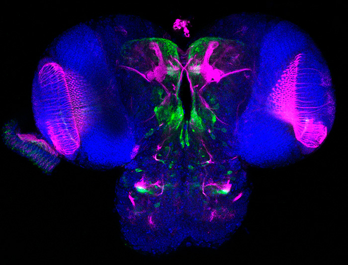
Part of this work was recognized in 2021 by the Chilean Society for Cellular Biology, which presented her with "The Best Image in Cellular and Molecular Biology” Award.
The challenge was to extract the brain and dissect it without damaging it. A task that took her two years. That photograph was taken during the pandemic, between lockdowns, and was part of the experiments that led to the scientific publication.
This new discovery reinforced her interest in continuing to work on the subject.
“It was very entertaining to spend hours watching, studying, and learning about flies. It was challenging, and that’s the fun part. I also really enjoyed having insight into both labs and being able to help my colleagues answer their questions. I am looking forward to having another model to test molecular targets in an accessible way in the future. How? I believe Professors Campusano and Marzolo said it best: We have to break down walls and talk... open up science and support it," concluded Francisca Rojo.
Basic Science and Interdisciplinarity Were Key in This Discovery
This publication gives the Faculty of Biological Sciences a boost to continue its commitment to basic science, that is, science focused on the abstraction of the studied phenomena rather than on their immediate practical application and, above all, to support its researchers and to invest in human capital.
For the research team, it has been an enriching experience that needs to be recognized so that others will be encouraged to pursue interdisciplinary work.
“Having Dr. Rojo learn about cellular biology from my lab was amazing. Seeing images like the one recognized by the SBCCH was a long-held dream of ours. Undoubtedly, it is a team effort and this reflects the fact that one of the best assets we have in the Faculty is our human capital. We have great students, and we need to support them," said Professor Jorge Campusano enthusiastically.
"Interdisciplinarity is fundamental. It was a gamble and it worked out... with a lot of effort from both laboratories, since this project was not directly financed by any current fund. Despite this, it opened the doors to a new line of research, in which there are now two more students working collaboratively. This is basic science research that has biomedical applications. Unfortunately, basic science receives less funding in Chile. We need to make new investments to be able to conduct more of this type of research," added María Paz Marzolo.


